ELECTRICAL CONDUCTION OF THE HEART
MYOCARDIUM DEPOLARIZATION
- Phase 0: Initial upswing of action
potential.
- Na+ Channels open until threshold is
reached.
- Phase 1: The potential may
repolarize slightly before starting the plateau phase.
- Na+ Channels are inactivated.
- Outward Rectifier K+
Channels open transiently, causing slight repolarization.
- Membrane potential remains near zero.
- Phase 2: Plateau Phase
-- This stage is responsible for prolonging the cardiac action
potential, making it longer than a nerve action potential.
- Ca+2 Channels open,
to keep the cells depolarized.
- Phase 3: Repolarization
- Ca+2 Channels close.
- Delayed Rectifier K+
Channels open to effect normal repolarization.
- Phase 4: Diastolic membrane
potential.
- Inward Rectifier K+ Channels
(different than the ones above) are open, to maintain resting potential.
- They are open at highly negative membrane
potentials (i.e. hyperpolarization-activated).
SA-NODE DEPOLARIZATION: It is similar to
depolarization in the myocardium, except for the following differences:
- Depolarization results from influx of Ca+2
rather than Na+
- There is no plateau phase (no Phase 1 and 2).
- Automaticity: Hyperpolarization-activated
cation current is activated at low potentials, resulting in automaticity of
the SA-Node.
- Epinephrine increases the rate of rise and
acetylcholine decreases the rate of rise of Phase-4 depolarization.
REFRACTORY PERIOD: Cardiac muscle cells
have prolonged refractory periods, to prevent tetany of cardiac muscle.
AUTONOMIC REGULATION of HEARTBEAT:
- Acetylcholine slows heart rate by
increasing K+ permeability.
- Norepinephrine speeds heart rate
by increasing the rate of rise of the cardiac action potential
during phase 0.
PROPAGATION of ACTION POTENTIAL:
- ATRIAL CONTRACTION: It takes about
70 msec to get from the SA-Node ------> depolarize the atria ------> to the
AV-Node.
- AV-NODAL DELAY: There is a delay in depolarization
of about 90msec, once the impulse reaches the AV-Node.
- The function of this delay is to separate the
contraction of the atria (i.e. atrial systole) from that of the
ventricles (ventricular systole), so that more blood has a chance to
fill into the ventricles.
- The AV-Node depends on slow-conducting Ca+2
Channels for depolarization, which helps to explain its slow rate
of depolarization.
- A smaller cell-size also helps to explain the
slow rate of conductance.
- BUNDLE OF HIS
- BUNDLE-BRANCHES: Two continuing branches of the
Bundle of His.
- Left Bundle Branch: It
depolarizes first. Depolarization goes from the left side of the
ventricular septum to the right side, accounting for the Q-Wave.
- Right Bundle Branch: It
depolarizes after the left side.
- PURKINJE SYSTEM: Very fast conduction.
- VENTRICULAR MUSCLE
- As depolarization proceeds in the ventricles,
it moves from endocardium ------> epicardium.
EKG LIMB LEADS:
-
Depolarization
occurs toward the positive side (the positive sides are labelled to
the right, and the respective negative sides are unlabeled).
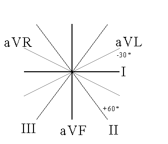
- HEXAXIAL SYSTEM: The positive end of each limb lead
is as follows:
- I: 0
- II: +60
- III: +120: In a normal ECG,
Lead III should have a net-zero QRS-Complex, as it is
perpendicular to aVR.
- aVR: -150: In a normal ECG,
the aVR lead should have a completely negative QRS Complex.
- aVL: -30
- aVF: +90
- DIRECTION OF ECG DEFLECTION: A positive deflection
on an ECG represents a depolarization that is traveling toward the positive
side of a particular lead.
- Maximal Positive Deflection: Occurs
when depolarization vector is in the exact same direction as the limb
lead.
- Zero net deflection: Occurs when
depolarization vector is exactly perpendicular to limb lead.
- Maximal Negative Deflection: Occurs
when depolarization vector is in the exact opposite direction as the
limb lead (i.e. in the direction of the negative end).
ELECTROCARDIOGRAM:
- P-WAVE: Atrial depolarization.
P-Wave duration is normally 80 msec.
- PR-INTERVAL: The distance from
the beginning of the P-Wave to the beginning of the Q-Wave.
- PR-Interval is the period from beginning of
atrial depolarization to the beginning of ventricular
depolarization.
- PR-Interval is normally 180-220 msec.
- PR-SEGMENT: The distance from
the end of the P-Wave and the beginning of the Q-Wave.
- QRS-COMPLEX: Ventricular
Depolarization. QRS Duration is normally 30-100 msec.
- Individual Components:
- Q-WAVE: Depolarization
of the septum. On most leads (except III and aVR) the Q-Wave
points downward if it can be seen at all. Septum
depolarization goes from the left side of the septum to the right
side.
- R-WAVE: Depolarization of
the ventricles. Sharp upward turn.
- S-WAVE: Return of
volt-potential to zero, because all the ventricular muscle has
depolarized and is therefore once again isoelectric.
- Sharp downward turn back to isoelectric
point. The S-Wave may go slightly negative before return back to
isoelectric point.
- QT-INTERVAL: From beginning of
Q-Wave to end of T-Wave. QT-Interval is normally 260-490 msec.
This is the period from beginning of ventricular depolarization to the
end of repolarization.
- ST-SEGMENT: Short segment from end of S-Wave to
beginning of T-Wave.
- ST-INTERVAL: From end of S-Wave to end of
T-Wave.
- RR-INTERVAL: Distance between
QRS-Complexes, or the distance between heart beats in a normal sinus
rhythm.
- T-WAVE: Repolarization of
Ventricles. Atrial repolarization masked by QRS-Complex.
- Repolarization occurs in the opposite direction
as depolarization, but the vector still points in the same direction
because the change in voltage also has an opposite sign.
- In the ventricles, the first tissue to
depolarize is the last tissue to repolarize.
READING THE ECG:
- Vertical Direction: 10 mm = 2 big boxes = 1 mV
deflection.
- Horizontal Direction:
- 1 mm = 40 msec.
- At standard speed, there are 25 mm, or 5 big
boxes, in each second.
- Speeds:
- Standard Speed = 25 mm/sec
- Extra-Sensitivity Speed = 50 msec, at which
point all values above must be doubled.
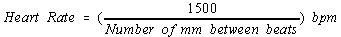
- Calculating Heart Rate Shortcut:
At standard speed:
PRECORDIAL LEADS: V1 thru V6 are placed to specific
places on the chest, for advanced ECG diagnostics. V1 is right-most, near the
SA-Node, while V6 is leftmost, past the apex of the heart.
MEAN ELECTRICAL AXIS OF THE HEART:
- Two ways to graphically determine mean electrical
axis:
- SHORT WAY: This is only accurate when there is
a net QRS-Deflection of virtually zero (i.e. the R deflection is equal
and opposite to the S deflection).
- Determine the lead that has a net zero
QRS-Deflection.
- On the hexaxial system, the mean electrical
axis points in the direction that is perpendicular to that lead.
- LONG WAY: This is longer but more accurate.
- Consider any two of the six hexaxial leads.
Determine again the Net QRS-Deflection for each lead.
- Plot that deflection along the appropriate
axis on a hexaxial chart.
- Draw a dotted line perpendicular to each of
the above plots, and extend the two lines until the intersect each
other.
- The Mean Electrical Axis is the vector that
points from the center to the intersection of those two lines.
- LAB: Different physiological effects on the mean
electrical axis:
- INSPIRATION: The diaphragm moves down ------>
It pulls the apex of the heart toward the right (i.e. in a more vertical
direction) ------> the mean electrical axis is more positive (+ more
degrees).
- FORCED EXPIRATION: The exact opposite of above.
The apex of the heart gets pushed upward and toward the left horizontal
axis ------> the mean electrical axis is less positive or even negative.
- PREGNANCY: The mean electrical axis would
deviate to the left, within normal limits. The physical presence of the
fetus would push up the diaphragm ------> heart leans toward left.
- LEFT VENTRICULAR HYPERTROPHY: Mean axis
deviation toward the left.
- Pulmonary Valve Stenosis: If we assume that it
leads to Right Ventricular Hypertrophy ------> Then we get (potentially
severe) right axis deviation.
- INFANCY: Right Axis Deviation, because the
infant's right ventricle and left ventricle musculature are about the
same size at birth. Left ventricle becomes larger within a couple
months.
- NORMAL MEAN AXIS: Anywhere between
-30 and +110.
- Anything negative of -30 is left axis
deviation, as occurs from left ventricular hypertrophy.
- Anything positive of +110 is right axis
deviation, as occurs from right ventricular hypertrophy.
ECG ABNORMALITIES:
- SINUS BRADYCARDIA: A heart rate
slower than 60 SA-Nodal depolarizations per minute. "Sinus" indicates that
the cardiac impulse is originating from the SA-Node as normal.
- SINUS TACHYCARDIA: Heart rate
faster than 100 bpm, originating as normal from the SA-Node.
- Tachycardia generally means you'll see a
shorter RR-Interval (i.e. faster heart rate).
- SINUS ARREST: No SA-Node
depolarization.
- This can be artificially induced by
carotid massage, which results in overstimulation of the Vagus
------> SA-Node hyperpolarized.
- ATRIAL PAROXYSMAL TACHYCARDIA:
Faster heart rate resulting from an ectopic pacemaker in
the atrial muscle.
- In the example the P-Wave points
downward because the atrial depolarization starts in the LA,
because that is where the tissue is leaky.
- BUNDLE-BRANCH BLOCKS: There is
some conduction block in the Bundle of His (Left or Right Bundle branches),
with results as below:
- 1 BLOCK: Partial block.
The PR-Interval is longer than normal
because it takes longer to conduct the impulse from SA-Node to AV-Node.
- 2 BLOCK: A QRS-Complex
occurs only after every other P-Wave. In other words, it takes two
P-Waves to sufficiently excite the AV-Node to conduct the impulse to the
ventricles.
- 3 BLOCK: There is no
temporal relationship between the P-Wave and QRS-Complex. Atrial
and ventricular depolarizations are being controlled by their own
independent pacemakers (the SA-Node and AV-Node respectively).
- AV-NODAL TACHYCARDIA: Tachycardia,
plus the P-Wave is insignificant or absent.
- This is tachycardia, where the impulse
originates from the AV-Node. The inherent pacemaker of the AV-Node is
faster than the SA-Node.
- PREMATURE VENTRICULAR CONTRACTION (PVC):
A premature QRS-Complex, or one that occurs without being preceded by a
P-Wave.
- That means that the P-Wave didn't start the
impulse, but it started somewhere else.
- Ectopic Pacemaker: With PVC,
the impulse originates in the ventricular muscle itself, due to leaky
membranes in the muscle.
- VENTRICULAR FIBRILLATION: Waves of
depolarization traveling in multiple directions all over the ventricular
muscle. The pacemaker activity is lost.
- ATRIAL FIBRILLATION: Fibrillation
in the atria is not serious in children, but it is serious in old people.
- That's because in old people, atrial systole
contributes a greater relative blood volume to cardiac output than in
children.
CLINICAL LECTURE: WOLF-PARKINSON-WHITE SYNDROME
- Normally, the AV-Node is the only pathway
for conduction of the impulse from the atria to the ventricles.
- Bachman's Bundle: Normally
conducts the impulse from Right Atrium to Left Atrium during atrial
systole.
- Moderator Band: Normally
conducts the impulse from the right ventricular septal wall to the right
free wall during ventricular systole.
- Lupus Erythematosus: Rare
condition associated with pediatric bradycardia. Usually pediatric heart
problems result in Tachycardia -- not bradycardia.
- PEDIATRIC TACHYCARDIAS: They are divided into two
types
- Supraventricular Tachycardia (SVT):
One where the problem originates somewhere in the AV-System.
- Ventricular Tachycardia (VT):
Problem originates in the ventricular system.
- Wolf-Parkinson-White Syndrome:
Extra conductive tissue in the myocardium, creating an accessory
pathway for conduction from atria to ventricles.
- This accessory pathway ultimately results in a
Reentry Tachycardia, or a conduction loop between the normal and
accessory pathways.
- The Wolf-Parkinson-White ECG:
Shorter PR-Interval due to rapid conduction of signal to ventricles
through accessory pathway.
- This is the ECG when the patient is
healthy and no problems are going on.
- The P-Wave and the QRS-Complex are
scrunched together, creating the appearance of a delta-wave
(hump right before QRS), and a longer overall QRS Complex.
- Reentry Tachycardia: You get it
from a unidirectional block in one pathway, coupled with slowed
conduction of an alternative pathway. This results in continuous
impulse conduction, or circus dysrhythmia.
- With WPW, the accessory pathway can get blocked
because it hasn't had the time to repolarize, then the normal pathway
provides a mean for retrograde conduction of depolarization.
- This results in a conduction loop and severe
tachycardia.
- TREATMENT: Slow down the conduction through one
pathway or the other.
- Use Ca+2-Channel Blockers (such as
Verapamil)
- Use Digoxin to increase
AV-Nodal sensitivity to ACh.
- Use beta-Blockers to block the normal NorE
sympathetic receptors on the AV-Node and cardiac muscle.
- In severe cases, surgically remove the
conductive tissue from the myocardium.
|